Research Methods
Physical genomics leverages the breakthrough computational modeling and super-resolution imaging and nanosensing techniques developed at CPGE.

Computational Modeling
Computational transcriptional modeling techniques developed at CPGE allow researchers to understand how gene transcription works at multiple interrelated levels, from individual DNA strands to the whole chromatin structure.
Imaging and Sensing Technologies
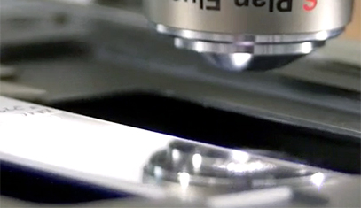
Novel super-resolution optical techniques developed by researchers at CPGE can image and sense structures across length scales, from individual DNA strands all the way up to thousands of live cells in real-time.
Novel Techniques
Partial Wave Spectroscopic (PWS) Microscopy
Partial Wave Spectroscopic (PWS) microscopy is an optical technique that uses light to understand what is happening at the nanoscale within cells. PWS allows researchers to examine chromatin structure in living cells, in real time, at the 20-200 nanometer length scale which, for example, is exactly where chromatin undergoes a transformation when cancer risk begins to increase. Not only is the technique label-free, allowing researchers to study chromatin within unaltered, living cells, but it does so with high-throughput and at very low cost.
Spectroscopic Intrinsic-contrast Photon-localization Optical Nanoscopy (SICLON)
SICLON is based on a recently discovered physical effect: biopolymers such as DNA, that until recently had been considered “dark” in the visible spectral range, fluoresce when illuminated by low-intensity visible light, a phenomenon which can be used for label-free nanoscopic imaging. SICLON combines time-resolved stochastic photon localization with simultaneous spectroscopic molecular-signature-carrying intrinsic fluorescence detection.
SICLON couples the principles of single-molecule nanoscopy with two key innovations: the endogenous fluorescence photoswitching effect, which bypasses fluorescence labeling, and a new technology able to simultaneously record not only the positions of emitting molecules but also their optical spectra, analysis of which enables SICLON to achieve molecular recognition and spatial resolution down to a few nanometers. SICLON is currently able to image unmodified DNA and chromatin without labeling with 6 nm spatial resolution, with work underway by CPGE researchers to improve this resolution further.
Inverse Spectroscopic Optical Coherence Tomography (ISOCT)
ISOCT is an emerging technology that combines the 3D spatial resolution of OCT (~1-5 µm resolution and hundreds of microns depth of imaging) with spectral analysis of the signal from each resolution voxel to measure the statistics of subdiffractional macromolecular density arrangement inside the voxel, with sensitivity down to 30 nm length scales. In particular, ISOCT is able to measure the scaling of chromatin packing density in cells located hundreds of microns below the tissue surface as part of complex 3D tissue models (e.g. organoids, matrix models) or in situ / in vivo. The power of ISOCT lays in its capacity for fast (~1mm3/sec) in situ and in vivo imaging of tissue in 3D with micron resolution and nanoscale sensitivity, utilization of low light power, and enabling of fiber-optic probe implementation for in vivo use.
Back to top
