Resources
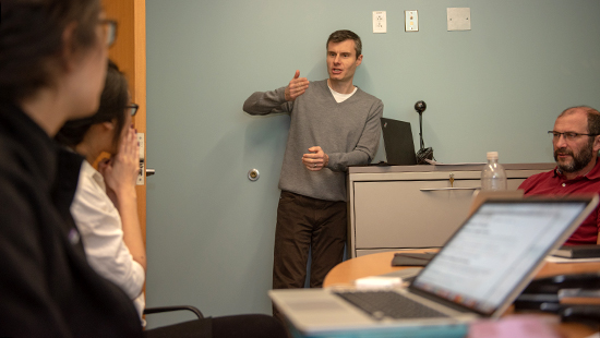
The Center for Physical Genomics and Engineering develops new, cutting-edge instrumentation and offers its investigators use and training on a wide array of unique imaging equipment and core facilities, providing access to state-of-the-art technology spanning all disciplines from engineering to biology, chemistry, physics, and medicine. Learn more about Northwestern's Core and Shared Facilities.
Novel Biophotonics Software and Instrumentation
The instruments at the CPGE are capable of imaging and sensing cellular structures at the nanoscale.
ANGORA
Angora is a free, open-source software package that computes numerical solutions to electromagnetic radiation and scattering problems. It is based on the finite-difference time-domain (FDTD) method, which is one of the most popular approaches for solving Maxwell's electrodynamics equations.
Learn more
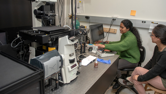
Confocal PWS
This system allows the acquisition of both PWS and confocal images and their combination in nearly real time by using a specially designed image processing suite.
AOTF/Spectrometer Benchtop PWS System Prototype
This system is designed to take PWS measurements of fixed cells and tissue samples and has been used extensively for clinical experiments evaluating PWS as a method for lung cancer screening and studies on prostate cancer progression risk assessment.
The system combines two methods of spectral filtration in a custom-built, automated benchtop microscope.
An acousto-optic tunable filter (AOTF) arm provides high-speed spectrally tunable illumination, while images are collected with a high speed CMOS camera and a secondary optical path allows high-resolution spectral sampling with a spectrometer and CCD camera scanned over the microscope image.
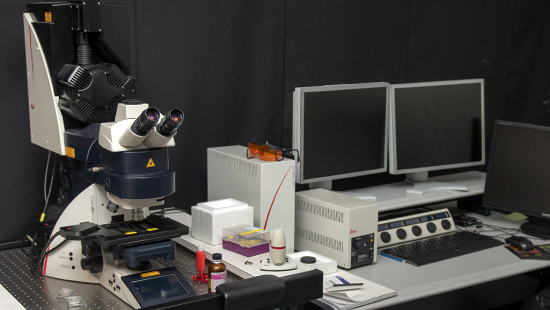
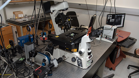
Live-Cell PWS Microscope
This system enables high contrast images of label-free live cells to be obtained through PWS spectroscopic analysis which is sensitive to the organization of nanoscale structure. By studying live cells, this system can perform time-dependent causation studies whereas PWS cytology is limited to studying correlations between populations at fixed time points.
Due to its configuration the live-cell PWS instrument is capable of multi-modal acquisition, including wide-field fluorescence and phase contrast microscopy, while maintaining physiological conditions for live cells thus enabling long-term experiments. The system allows multiple cells (1-100 depending on the eukaryotic cell line and magnification) to be imaged simultaneously with the acquisition of the full interference spectra for analysis taking under 30 seconds.
High-Throughput Live-Cell PWS Microscope
The high-throughput live-cell PWS microscope is intended to expand the capabilities of the live-cell PWS technology. With this microscope multimodal colocalization experiments can be performed with wide-field fluorescence and phase contrast in addition to long-term time lapse experiments with very high temporal resolution capable of resolving dynamic real-time changes in nanoscale cellular structure. The microscope is equipped with an automated filter turret and sample stage as well as an incubation chamber and focus maintenance system for time lapse experiments and enables PWS images to be acquired at up to 100 frames per second, allowing imaging of the dynamics of nanoscale structure within live cells.
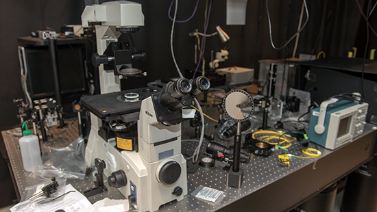
Spectroscopic Intrinsic Contrast Photolocalization Optical Nanoscopy (SICLON)
The spectroscopic single-molecule localization microscope is designed to enable spectroscopic super-resolution imaging of cells with the ability to co-register these images to PWS images of nanoscale structure. The system is able to image unmodified DNA and chromatin without labeling with 6 nm spatial resolution and incorporates automated sample stages, filter turrets, a live-cell incubator, a focus maintenance system, a motorized laser illumination system allowing TIRF illumination, and high NA objectives for high-resolution imaging. The system includes several Illumination sources: a white-light LED for widefield fluorescence and PWS and a selection of lasers for TIRF/single molecule localization experiments. Spectral filtration for PWS is provided by a liquid crystal tunable filter, enabling automated wavelength tuning. Images are acquired with one of three cameras allowing various specific modalities of spectroscopic imaging. One of the imaging ports is coupled to a custom-made spectrometer and camera which enables spectroscopic single-molecule localization experiments and simultaneous imaging of multiple fluorophores. Modalities this system can perform include: super-resolution single-molecule localization fluorescence, spectroscopic super-resolution single-molecule localization fluorescence, super-resolution single molecule localization of intrinsic fluorescence, multidimensional widefield fluorescence (x,y,z,λ,t), fixed/live-cell PWS, TIRF, and phase contrast.
Optical Coherence Microscope (OCM)
The optical coherence microscope is an optical coherence tomography (OCT) system built into an inverted microscope. It is a Fourier-domain OCT system that uses a custom built confocal scanner with two galvo mirrors and visible light spectrometer to sample interference spectra from each pixel in the scanned image region. The system enables label-free 3D imaging of cells and tissue at microscopic resolutions. Due to its optical design and incorporation into a commercial microscope body, this system can be made multimodal in the future to perform custom confocal fluorescence experiments or PWS.
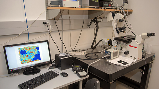
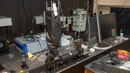
Scanning PWS System
The scanning PWS system aims to significantly reduce acquisition time for the PWS system. The equipment has two major components: a microscope imaging system and a Michelson interferometer system. The sample forms an intermediate image that is collimated into an interferometer. The light coming out of the interferometer system is collected by a lens and imaged onto a CCD camera. As the PZT stage scans the interference range, a series of images are acquired. The time series data for each point in the images is inverse-transformed to Fourier domain so that the spectral information can be obtained. The spectral data can be processed in the same way as in the traditional PWS system to measure the nanoscale statistical properties of samples. The acquisition time for a spectrum cube can be reduced to less than one second.
D3 ISOCT Benchtop System
This system aims to obtain general morphology, angiography, blood oxygenation, and nanoscale characterization in living specimens. The system utilizes several light sources allowing collection in the visible and infrared range. Two bands are used to allow for decreased variance in nanoscale characterization: the infrared band allows for deeper imaging of morphology and angiography measurements, and the visible band increases sensitivity to blood oxygenation measurements.
Additionally, a miniaturized D3 ISOCT probe was developed (2 mm diameter). The probe allows for in vivo measurements in locations not accessible by the benchtop system, such as the colon.